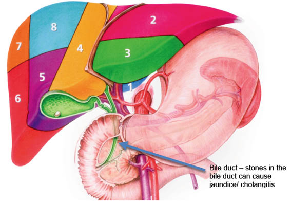
What are bile duct stones?
The liver makes bile, which is used to digest fats. The bile duct carries bile from the liver to the duodenum where digestion of fat begins. The normal width of the bile duct is 3-5mm. Bile duct stones can occur in approximately 10-15% of patients with gallstones, but only become symptomatic in 1-4% of patients. Importantly bile duct stones can form in patients even after the gallbladder has been removed.
Who commonly presents with bile duct stones?
Any patient with gallstones can develop bile duct stones.
How do bile duct stones present?
Bile duct stones can present with symptoms similar to gallstones such as pain in the upper abdomen and nausea especially after consumption of rich or fatty food. Bile duct stones can obstruct/ block the flow of bile into the small bowel/ duodenum, which presents with Jaundice. Patients commonly present with yellow discolouration of their skin, dark urine and pale stools. In severe cases jaundice is associated with a fever and pain, this condition is known as cholangitis and is treated as a surgical emergency with strong pain killers, antibiotics and an ERCP.
Bile duct stones can also present with Acute Pancreatitis. Small gallstones, biliary crystals or 'sludge' can pass into the bile duct and block the main pancreatic duct resulting in acute pancreatitis. This is a condition that usually presents with severe abdominal pain, nausea and vomiting. Pancreatitis tends to be treated in hospital with painkillers and fluids to allow the symptoms to settle, following which, the gallbladder is removed, by keyhole surgery at the earliest opportunity. In mild case of acute pancreatitis if the symptoms settle, the gallbladder is removed on the same admission.
How are bile duct stones diagnosed?
Ultrasound: The most sensitive test performed when gallstones are suspected is the ultrasound examination. Ultrasound machine utilises sound waves to create images of the gallstones and other organs. The sound waves from the machine bounce off the gallstones and other organs and the images are captured on a video monitor. If the bile duct contains stones, the bile duct itself can be dilated to at least 1cm or more.
Magnetic resonance cholangiogram (MR scan): is used to diagnose suspected bile duct stones. It is non-invasive and is used when the suspicion of bile duct stone is high.
Endoscopic Ultrasound (EUS): can be used to diagnose stones from the bile duct as well as biliary sludge. It involves passing a flexible camera tube down the throat into the small bowel (duodenum). The flexible camera has an ultrasound probe at its tip, which can accurately diagnose both stones in the gallbladder and bile ducts. It is sometimes combined with an ERCP, which is designed to remove bile duct stones.
Computed tomogram (CT/ CAT scan): sometimes is used mainly in the evaluation of complications of bile duct stones.
How are bile duct stones treated?
Fifty percent, 50%, of bile duct stones pass spontaneously into the small bowel/ duodenum and require no intervention. Once symptomatic, giving rise to either jaundice, pain, cholangitis or pancreatitis, in some cases a combination, they can be treated by;
1) ERCP or
2) Laparoscopic bile duct exploration
The laparoscopic 'keyhole' route is only possible in some patients with most surgeons reporting a success rate of 40-60%. This procedure is combined in most circumstances along with gallbladder surgey.
Very rarely if the above two treatments have failed in some cases open surgery is required to remove the bile duct stones and possibly reconstruct the bile duct with a loop of small bowel, Hepaticojejunostomy or Choledochojejunostomy.